Spinal Diagnostics
Despite the fact that many causes of neck and back pain can be diagnosed with a thorough history and physical examination, additional testing such as x-ray, computed tomography (CT), magnetic resonance imaging (MRI), and electrodiagnostic testing (EMG/NCS) is often employed to more conclusively and precisely determine a patient’s primary pain generating structure. However, in some cases of neck and back pain, diagnostic injections remain the gold standard to best identify the main cause of a patient’s spine-related pain.

Selective Nerve Root Block
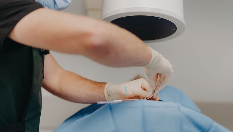
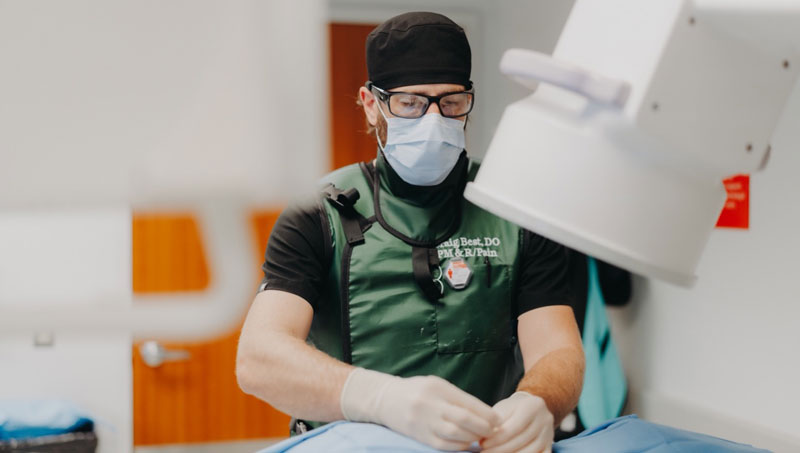
Selective nerve root blocks can be utilized to help determine if a particular nerve root is the primary source of a patient’s pain. Using x-ray (fluoroscopic) guidance, a needle is placed near the suspected painful nerve root. Then, a small volume of numbing medication (local anesthetic) is administered to temporarily block the pain coming from the target nerve. If a significant decrease in pain is noted during the time period the local anesthetic is acting on the nerve, it can then be determined with increased certainty that the targeted nerve is the primary source of pain.
Medial Branch Blocks
When a patient’s facet (zygapophysial) joints are emanating pain, the medial branch nerves will sense this pain signal and transmit it along a pathway to the spinal cord and up to the brain where we interpret and “feel” that painful sensation. As such, medial branch nerve blocks can be used as a diagnostic injection to help determine whether or not the facet joints are the primary source of pain. Using x-ray (fluoroscopic) guidance, needles are safely guided to and placed near the medial branch nerves which are then anesthetized, thus temporarily blocking the pain coming from the facet joints. Patients are then asked to monitor their pain during the time period that the local anesthetic is blocking the facet joint pain. In patients who experience a significant improvement in their pain, it increases the likelihood that the facet joints are the main source of pain and radiofrequency ablation can then be performed as a treatment to provide long lasting improvement in the pain. In patients who do not experience much improvement in their pain during the anesthetic phase, it can be determined with much more certainty that the facet joints are not the main source of the pain and the focus can shift to other potential pain generating structures.

Sacroiliac Joint Injection
Sacroiliac (SI) joint pain typically causes low back/buttock region pain that is reproduced with certain provocative maneuvers during the physical examination. Ultimately, patient response to SI joint injection remains the gold standard diagnostic test to best determine whether or not the SI joint is the primary cause of a patient’s low back pain. Using x-ray (fluoroscopic) guidance, a needle is guided into the SI joint. Once the needle is in the joint, numbing medication (local anesthetic) is instilled through the needle to anesthetize the SI joint. If the patient experiences a significant improvement in pain during the time the local anesthetic is blocking the painful joint, it can be determined with increased certainty that the SI joint is the primary source of pain.
Sacral Lateral Branch Blocks
When a person’s sacroiliac (SI) joint is emanating pain, the sacral lateral branch nerves will sense this pain signal and transmit it along a pathway to the spinal cord and up to the brain where we interpret and “feel” that painful sensation. As such, lateral branch nerve blocks can be used as a diagnostic injection to help determine whether or not the SI joint is the primary source of pain. Using x-ray (fluoroscopic) guidance, needles are safely guided to and placed near the lateral branch nerves which are then anesthetized, thus temporarily blocking the pain coming from the SI joint. Patients are then asked to monitor their pain during the time period that the local anesthetic is blocking the SI joint pain. In patients who experience a significant improvement in their pain, it increases the likelihood that the SI joint is the main source of pain and radiofrequency ablation can then be performed as a treatment to provide long lasting improvement in the pain. In patients who do not experience much improvement in their pain during the anesthetic phase, it can be determined with much more certainty that the SI joint is not the main source of the pain and the focus can shift to other potential pain generating structures.

Provocative Lumbar Discography
Provocative lumbar discography is the gold standard diagnostic test to precisely confirm or exclude the lumbar intervertebral disc as a source of chronic low back pain. Using fluoroscopic (x-ray) guidance, needles are placed within two or more discs and pressurized using contrast dye. Based on patient response (i.e. whether the patient’s typical low back pain is reproduced or not) and imaging findings such as fissures (tears) within the disc(s), a diagnosis of lumbar discogenic pain (discogenic low back pain) can be confirmed or ruled out as a source of chronic low back pain.
Patients who have been experiencing low back pain for greater than 3 months, have tried and failed conservative management (medications, physical therapy, chiropractic), and for whom non-invasive diagnostic tests (x-ray, MRI) have failed to precisely diagnose the source of low back pain.
Low back pain is a very common cause of musculoskeletal disability, but “low back pain” is not a diagnosis. There are distinct anatomic spinal structures that can potentially generate pain. Studies have shown that the intervertebral discs are the main pain generating structure in 40-70% of cases of axial (non-radiating) low back pain. In patients younger than 60 years of age, the disc is the most common cause of low back pain, followed by the sacroiliac joint then the facet joint. In patients older than 60 years of age, the facet joint is the most cause of low back pain, followed by the disc then the sacroiliac joint. As such, provocative lumbar discography is an important tool to precisely diagnose pain emanating from the lumbar intervertebral disc(s).
Patients with discogenic low back pain typically complain of deep and aching pain that can become sharp with movement, an inability to sit for prolonged periods of time, temporary relief with change of position, and low back more than leg pain. This type of low back pain commonly begins with a lifting and/or twisting injury.
Despite concerns raised by some physicians that lumbar discography contributes to higher rates of disc degeneration, a recent review of the safety and overall diagnostic value of provocative lumbar discography concluded that it is a safe and helpful diagnostic test when performed by properly trained physicians using strict procedural guidelines as outlined by the Spine Intervention Society.
First, in patients who eventually go on to have lumbar fusion surgery for management of their discogenic low back pain, there is an 88% chance of success with a positive discogram compared to just a 50% chance of success with a negative discogram. Second, when lumbar discography identifies the source of a patient’s low back pain earlier in their clinical course, it can save the patient from potentially excessive and unnecessary testing and treatment. Lastly, it can potentially provide peace of mind by having a precise diagnosis of one’s low back pain.
At a Glance
Dr. Craig Best
- Harvard Fellowship-Trained Interventional Spine & Sports Medicine Specialist
- Double Board-Certified in Physical Medicnie & Rehabilitation and Pain Medicine
- Assistant Professor of Physical Medicine & Rehabilitation and Orthopedic Surgery
- Learn more

