Patellar Tendinopathy
What is patellar tendinopathy?
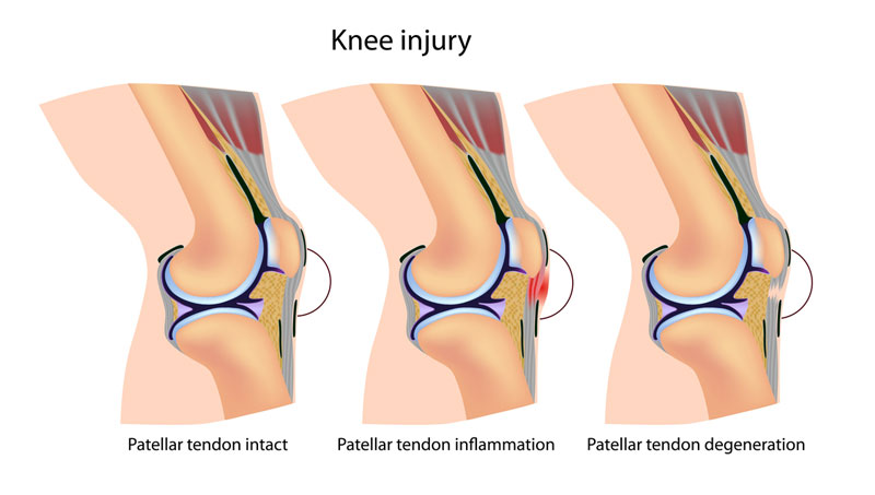
The quadriceps muscles transition to the quadriceps tendon and then into the patellar tendon as it transitions from the patella (knee cap) to the tibial tuberosity (bony prominence at the shin bone). When the patellar tendon becomes overloaded such as those seen in sports requiring repetitive jumping (basketball, volleyball, etc.), it may become inflamed and painful. This is termed patellar tendinitis. If the issue is not managed properly initially, this may progress to microscopic tearing and disorganization of the tendon fibers with ongoing pain and dysfunction. This is called patellar tendinosis.
How is patellar tendinopathy diagnosed?
Patients with patellar tendinopathy (tendinitis or tendinosis) present with anterior (front) knee pain that may be worsened jumping, walking up or down stairs, or squatting. Patients often feel transiently better after they’ve warmed up for their respective sport or exercise activity but note a return of the pain once they’ve cooled down. Physical examination may reveal tenderness along the patellar tendon and at the bony attachments, focal swelling at the tendon, and reproduction of pain with deep squats or resisted knee extension. X-ray, ultrasound, or magnetic resonance imaging (MRI) may aid in confirmation of diagnosis.
How is patellar tendinopathy treated?
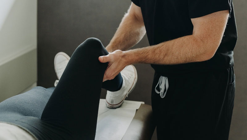
Initial treatment options for management of patellar tendinopathy may include medications and physical therapy. Physical therapy should primarily focus on hip, glute, quadriceps, and hamstring strengthening as well as overall load/volume management of exercise and training. If a patient is still experiencing significant pain despite the aforementioned treatment options, interventional options may include corticosteroid (cortisone) or platelet rich plasma (PRP) injections.
Corticosteroid Injection
Using ultrasound, a needle is carefully and precisely guided to the painful region. A steroid solution is then instilled through the needle to coat or bathe the painful region with anti-inflammatory medication. This helps to decrease inflammation and, subsequently, decreases pain and improves function.
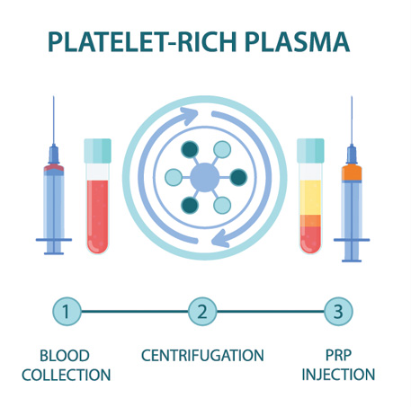
Platelet Rich Plasma (PRP)
PRP is component of the patient’s own blood. It is rich in growth factors and other cells that signal an increased healing response to a damaged tissue. It is used to treat a variety of painful spine and musculoskeletal conditions.
Blood is drawn from a patient and then placed in a centrifuge for it to be “spun down.” This causes the different components of the blood to separate out in the vial. The PRP solution is then drawn up into a syringe and prepared to be injected at the site of the patient’s injury.
Discontinue use of all non-steroidal anti-inflammatory drugs (NSAIDs) at least 7 days prior to the procedure. These may include ibuprofen (Advil, Motrin), naproxen (Aleve), meloxicam (Mobic), diclofenac (Voltaren), indomethacin (Indocin), and celecoxib (Celebrex). If you are taking oral corticosteroids such as prednisone or a Medrol Dosepak, please discuss this with Dr. Best prior to your procedure. In some cases, Dr. Best may request that the corticosteroid medication be discontinued in preparation for the PRP injection. Do NOT stop aspirin unless specifically instructed by Dr. Best. Depending which body part is injected, you may need a driver to and from your procedure. If you have any questions or concerns about whether to continue or discontinue any of your medications leading up to your PRP injection, please discuss these issues with Dr. Best and his team.
Once the PRP solution is created, the patient is positioned for the procedure. The skin is thoroughly cleaned and the target for the injection obtained with ultrasound or fluoroscopy (x-ray). Then, a numbing solution is injected at the skin and subcutaneous tissues for increased procedural comfort. Finally, under ultrasound or fluoroscopic (x-ray) guidance, the needle is guided to the injury site and the PRP solution is deposited.
It is common to experience mild to moderate pain or discomfort during the initial 0-3 days after the PRP procedure. Post-procedure pain can be easily managed with acetaminophen (Tylenol) or other non-NSAID pain medication. Try to avoid applying ice or heat to the injection site.
During the 3–14-day period after the PRP injection, you may gradually increase physical activity. Please continue to avoid use of NSAIDs; however, ice may be applied for short periods of time throughout the day to aid in management of post-procedure soreness/discomfort if present.
During the 2–4-week period after the PRP, Dr. Best may recommend initiation of a course of physical therapy to aid in recovery and optimization of healing. The patient may begin to note improvement in pain during this time period, though it often takes 1 month or more for the benefits of PRP to take hold.
At this time, PRP injections are not typically covered by any insurance companies. Pricing and payment options can be discussed with Dr. Best and his team prior to your procedure.
As an alternative to PRP injections, patellar tendon region injections with corticosteroid can be performed to help alleviate knee region pain. Using ultrasound, a needle is carefully and precisely guided to the painful region. A steroid solution is then instilled through the needle to coat or bathe the painful region with anti-inflammatory medication. This helps to decrease inflammation and, subsequently, decreases pain and improves function.
At a Glance
Dr. Craig Best
- Harvard Fellowship-Trained Interventional Spine & Sports Medicine Specialist
- Double Board-Certified in Physical Medicnie & Rehabilitation and Pain Medicine
- Assistant Professor of Physical Medicine & Rehabilitation and Orthopedic Surgery
- Learn more
