Rotator Cuff Injury
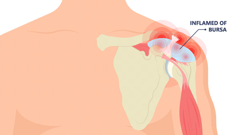
What is subacromial bursitis/impingement?
The subacromial space is delineated by the humeral head inferiorly (below) as well as the acromion, coracoacromial ligament, and acromioclavicular (AC) joint superiorly (above). Rotator cuff tendons, long head of the biceps tendon, subacromial bursa, and coracoacromial ligament are situated between the two aforementioned borders. Any factors that disturb or irritate the structures in the subacromial space may lead to pain and impingement.
What is rotator cuff tendinopathy?
There are four rotator cuff muscles and their respective tendons: supraspinatus, infraspinatus, teres minor, and subscapularis. Their primary role is to stabilize the shoulder joint during dynamic movement. Rotator cuff tendinopathy occurs when one or more of the rotator cuff tendons becomes irritated, inflamed, injured, and/or torn, leading to shoulder pain and dysfunction.
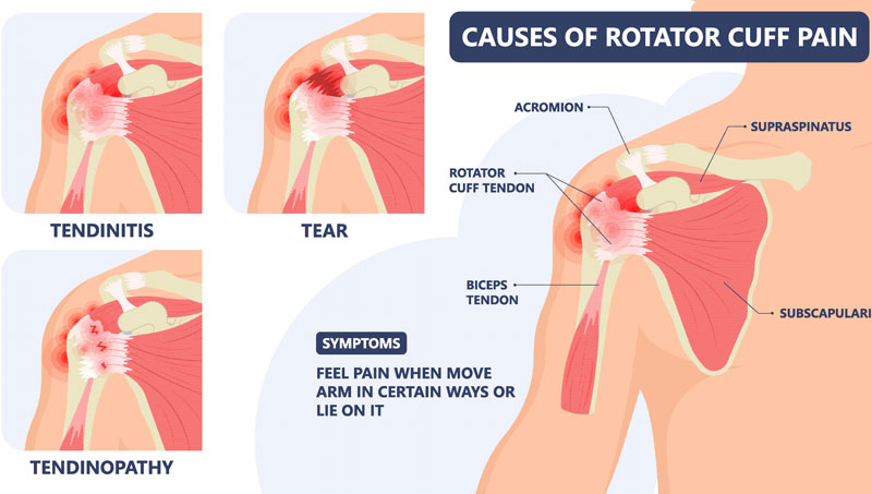
What causes subacromial bursitis/impingement and rotator cuff tendinopathy?
Many factors may lead to the development of subacromial bursitis/impingement and/or rotator cuff tendinopathy. Bony abnormalities such as the morphology of the acromion or AC joint arthritic changes may narrow the subacromial space and contribute to rotator cuff tendon injury. Abnormalities of glenohumeral and/or scapulothoracic biomechanics including the musculature that supports these joints may increase likelihood of developing subacromial bursitis/impingement and rotator cuff tendinopathy. Increased thoracic kyphosis (flexion) may also negatively impact shoulder biomechanics, resulting in pathologic changes at the subacromial space and rotator cuff tendons. Weakness and/or dysfunction of rotator cuff musculature contributes to poor shoulder biomechanics and predisposition to shoulder issues. Degenerative changes with aging may contribute rotator cuff tendinopathy. Finally, trauma, sports injuries, or occupational injuries may lead to acute or chronic rotator cuff pathology.
How is subacromial bursitis/impingement and rotator cuff tendinopathy diagnosed?
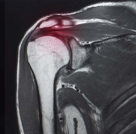
Patients with subacromial bursitis/impingement and rotator cuff tendinopathy typically present with anterolateral (front and side) shoulder pain that may be referred to the lateral (side) upper arm. The pain may be worsened with overhead activities or laying on the affected side. Physical examination may demonstrate painful shoulder range of motion, weakness of certain shoulder movements, and positive impingement signs and other provocative tests. X-ray, ultrasound, or magnetic resonance imaging (MRI) may aid in confirmation of diagnosis.
How is subacromial bursitis/impingement and rotator cuff tendinopathy treated?
Initial treatment options for management of subacromial bursitis/impingement and rotator cuff tendinopathy may include medications and physical therapy. Physical therapy should primarily focus on improvements in posture, shoulder region biomechanics, range of motion, and multi-planar shoulder and scapular region strengthening. If a patient is still experiencing significant pain and dysfunction despite the aforementioned treatment options, interventional options may include corticosteroid or platelet rich plasma (PRP) injections. Finally, referral to orthopedic surgeon skilled in shoulder arthroscopy/sports medicine may be considered for further evaluation and consideration of surgical intervention.
Subacromial Shoulder Injection
Using ultrasound, a needle is carefully and precisely guided to the subacromial space. Once the subacromial space has been accessed, a steroid solution is instilled through the needle to coat or bathe the painful region with anti-inflammatory medication. This helps to decrease inflammation and, subsequently, decreases pain and improves function.
Platelet Rich Plasma (PRP)
PRP is component of the patient’s own blood. It is rich in growth factors and other cells that signal an increased healing response to a damaged tissue. It is used to treat a variety of painful spine and musculoskeletal conditions.
Blood is drawn from a patient and then placed in a centrifuge for it to be “spun down.” This causes the different components of the blood to separate out in the vial. The PRP solution is then drawn up into a syringe and prepared to be injected at the site of the patient’s injury.
Discontinue use of all non-steroidal anti-inflammatory drugs (NSAIDs) at least 7 days prior to the procedure. These may include ibuprofen (Advil, Motrin), naproxen (Aleve), meloxicam (Mobic), diclofenac (Voltaren), indomethacin (Indocin), and celecoxib (Celebrex). If you are taking oral corticosteroids such as prednisone or a Medrol Dosepak, please discuss this with Dr. Best prior to your procedure. In some cases, Dr. Best may request that the corticosteroid medication be discontinued in preparation for the PRP injection. Do NOT stop aspirin unless specifically instructed by Dr. Best. Depending which body part is injected, you may need a driver to and from your procedure. If you have any questions or concerns about whether to continue or discontinue any of your medications leading up to your PRP injection, please discuss these issues with Dr. Best and his team.
Once the PRP solution is created, the patient is positioned for the procedure. The skin is thoroughly cleaned and the target for the injection obtained with ultrasound or fluoroscopy (x-ray). Then, a numbing solution is injected at the skin and subcutaneous tissues for increased procedural comfort. Finally, under ultrasound or fluoroscopic (x-ray) guidance, the needle is guided to the injury site and the PRP solution is deposited.
It is common to experience mild to moderate pain or discomfort during the initial 0-3 days after the PRP procedure. Post-procedure pain can be easily managed with acetaminophen (Tylenol) or other non-NSAID pain medication. Try to avoid applying ice or heat to the injection site.
During the 3–14-day period after the PRP injection, you may gradually increase physical activity. Please continue to avoid use of NSAIDs; however, ice may be applied for short periods of time throughout the day to aid in management of post-procedure soreness/discomfort if present.
During the 2–4-week period after the PRP, Dr. Best may recommend initiation of a course of physical therapy to aid in recovery and optimization of healing. The patient may begin to note improvement in pain during this time period, though it often takes 1 month or more for the benefits of PRP to take hold.
At this time, PRP injections are not typically covered by any insurance companies. Pricing and payment options can be discussed with Dr. Best and his team prior to your procedure.
As an alternative to PRP injections, subacromial shoulder injections with corticosteroid can be performed to help alleviate shoulder region pain. Using ultrasound, a needle is carefully and precisely guided to the subacromial space. Once the subacromial space has been accessed, a steroid solution is instilled through the needle to coat or bathe the painful region with anti-inflammatory medication. This helps to decrease inflammation and, subsequently, decreases pain and improves function.
At a Glance
Dr. Craig Best
- Harvard Fellowship-Trained Interventional Spine & Sports Medicine Specialist
- Double Board-Certified in Physical Medicnie & Rehabilitation and Pain Medicine
- Assistant Professor of Physical Medicine & Rehabilitation and Orthopedic Surgery
- Learn more
