Vertebral Compression Fracture
What is a vertebral compression fracture?
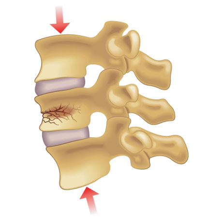
The vertebral body is the block shaped portion that makes up the anterior (front) aspect of a segment of the bony spinal column. The superior (top) and inferior (bottom) borders of the vertebral body are called the endplates. When a vertebral compression fracture (VCF) occurs, the superior and/or inferior endplate of the vertebral body is fractured which can be quite painful.
What causes a vertebral compression fracture?
The two main causes of VCFs include osteoporosis and malignancy. With osteoporosis, the bone mineral density is significantly decreased (i.e. thinning of bones) and predisposes a person to incur a fracture, typically with very little force. Similarly, malignancies can weaken the vertebral body and increase likelihood of developing a VCF. Finally, traumatic injuries such as a fall off of a ladder can cause a VCF.
How is a vertebral compression fracture diagnosed?
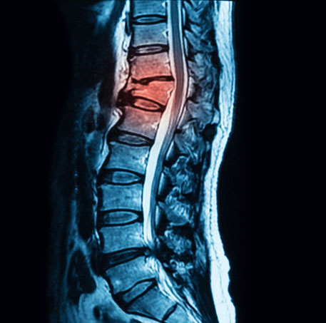
Patients with VCFs typically complain of significant midline axial low back pain that is worsened with lumbar flexion motions such as sitting or bending forward. Physical examination may reveal significant midline spinal tenderness and pain with light percussion over the painful vertebral segment(s). Further evaluation with x-ray, MRI, CT, and/or bone scan is warranted and reveals the VCF.
How is a vertebral compression fracture treated?
Initial treatment includes management of the pain with analgesic medications and bracing. In cases of severe pain, debility, and functional impairment when the VCF is in the acute or subacute stages, a procedure called a kyphoplasty may be performed to help stabilize the fracture and decrease pain. In addition, in cases of osteoporotic VCFs, patients may be referred to endocrinology for further evaluation and optimization of bone mineral density to hopefully decrease the risk of future osteoporotic fractures.
Kyphoplasty
Using x-ray (fluoroscopy), the VCF is identified. Local anesthetic is then utilized to numb up the tract from the skin and subcutaneous tissues all the way down to the bone, called the pedicle. A larger needle, called a trocar, is then advanced to the vertebral body through the pedicle. A balloon is then inserted through the trocar and into the vertebral body. It is then inflated in order to open up space which is then filled with medical grade cement. This helps to stabilize the VCF, which subsequently decreases pain and improves overall functional status.
At a Glance
Dr. Craig Best
- Harvard Fellowship-Trained Interventional Spine & Sports Medicine Specialist
- Double Board-Certified in Physical Medicnie & Rehabilitation and Pain Medicine
- Assistant Professor of Physical Medicine & Rehabilitation and Orthopedic Surgery
- Learn more
